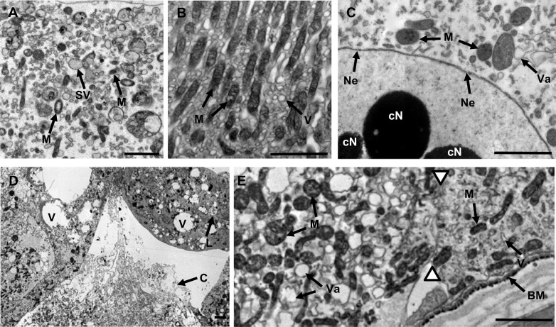
Fig 2.
Transmission electron micrographs of ultrathin sections (stained with 1% uranyl acetate) of midgut tissue from third-instar C. zealandica larvae 24 h following oral ingestion of either control treatment (TBS buffer only) (A and B) or purified Yen-Tc toxin (C to E). (A) The apical portion of a healthy active midgut cell showing structures involved in secretion and absorption. (B) The basal portion of a healthy midgut cell showing elongated mitochondria oriented along a basal-apical axis, surrounded by small smooth vesicles. (C) At 24 h post-toxin treatment, the region around the nucleus had variable-sized, irregular-shaped vesicles and swollen mitochondria with prominent dark spots, while the nucleus displayed a globular electron-dense condensation of nuclear material and evidence of breaks in the double membrane of the nuclear envelope. (D) Presence of both apoptotic-like cells (upper cell showing a condensed cytoplasm with evidence of organelles being broken down in lysosomes) and necrotic-like cells (lower cell showing cytoplasm escaping from broken membranes). (E) The two white triangles highlight the junction of two cells, revealing cell-to-cell variation, with the left-hand cell displaying variable-sized vesicles with irregular profiles and less-well-oriented mitochondria (both swollen and elongated mitochondria are present) and the right-hand cell showing less dramatic changes. Arrows point to highlighted structures labeled with as follows: B, basement membrane; cN, nucleus with condensed chromatin; M, mitochondria; Ne, nuclear envelope disrupted; SV, secretory vesicle; Va, vacuole. Paired white triangles indicate the boundary of two adjoining cells. Scale bars represent 2 μm.

