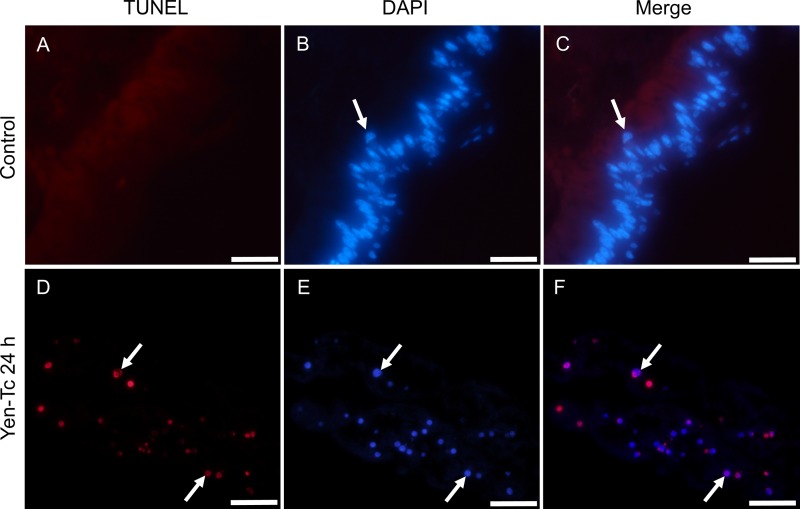
Fig 3.
TUNEL staining of longitudinal sections from third-instar C. zealandica larvae 24 h post-oral treatment with either the control (TBS buffer only) (A to C) or purified Yen-Tc (D to F). Arrows indicate examples of either TUNEL-positive nuclei (TMR label appears as red fluorescence) (A and D), nuclei stained with DAPI (blue fluorescence) (B and E), or nuclei displaying both TUNEL-positive and DAPI staining (purple appearance in merged images) (C and F). Scale bars represent 50 μm.

