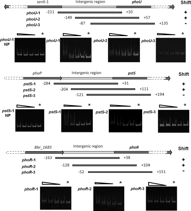
Fig 4.
PhoP binding assays to various promoter regions. Shown is a schematic representation of amplified DNA fragments used in gel mobility shift assays; the numbers correspond to the ends of the fragments relative to the presumed translation start site. Plus and minus signs indicate observed binding or no binding, respectively, of PhoP to specific sections of the phoU, pstS, or phoR promoter regions. The terms phoU-1-NP and pstS-1-NP represent binding assays with unphosphorylated PhoP, and the remainder of the bindings assays were performed using phosphorylated PhoP. PhoP was phosphorylated for 1 h at 25°C in the presence of acetyl phosphate. Each gel represents a binding assay with a particular DNA fragment as indicated in the left margin and decreasing amounts of (phosphorylated) PhoP (lanes 1 to 5 correspond to 400, 200, 100, 50, and 0 mM PhoP, respectively).

