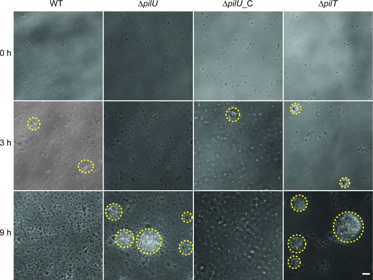
Fig 5.
Microcolony formation is delayed in a meningococcal pilU mutant in cell-free medium. Live-cell phase-contrast microscopy was performed on bacteria growing in cell-free medium. Representative images were selected based on when the wild type and the ΔpilU mutant reached their respective maximal microcolony sizes. Microcolonies are highlighted with a yellow dashed line around the periphery. The ΔpilT mutant was included as a positive control for aggregation. Bar = 10 μm.

