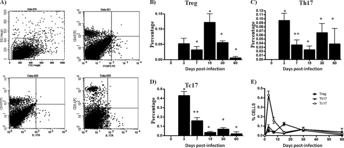
Fig 2.
Spleen T-cell dynamics during N. brasiliensis infection. The percentages of T-cell subpopulations were obtained using specific monoclonal antibodies and flow cytometry analysis. (A) Representative dot plots show the selected region, and specific staining was used to study the T-cell subpopulation. (B) CD4+ Foxp3+ (Treg) cell levels were increased at day 15 of infection. (C and D) CD4+ IL-17A+ (Th17) and CD8+ IL-17A+ (Tc17) cell counts were highest at day 3. (E) Dynamics of three subpopulations during N. brasiliensis infection. The data represent the means of five mice per group ± the SEM. Statistical analyses were performed using one-way ANOVA, and the post hoc test was Tukey's (*, P < 0.05; **, P < 0.01). The data presented in this figure are representative of three experiments of similar experimental design.

