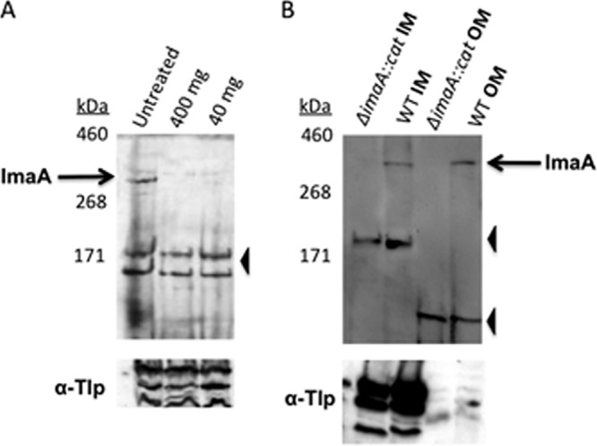
Fig 3.

ImaA localizes to the outer membrane. (A) Whole cells of H. pylori strain LSH100 were treated with different concentrations of proteinase K (40 or 400 mg ml−1) or with no proteinase K as a control. The top panels show blots probed with anti-ImaA-1, while the bottom panels are probed with anti-GST-TlpA22 antibody (α-Tlp), which recognizes inner membrane chemoreceptors (83). Similar results were obtained with strain mG27 (not shown). (B) Sarcosine-insoluble outer membrane (OM) fractions and sarcosine-soluble inner membrane (IM) fractions were obtained from wild-type (WT) and H. pylori SS1 ΔimaA::cat mutant cells and then probed with anti-ImaA-1. In both panels, the positions of full-length ImaA are indicated by black arrows labeled ImaA, and the positions of nonspecific proteins recognized by the anti-ImaA serum are indicated by black arrowheads.
