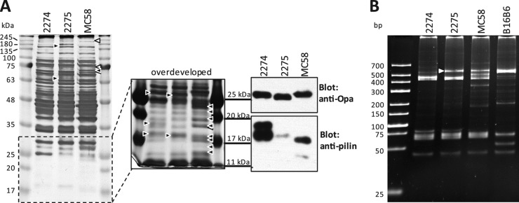
Fig 1.

Phenotypic characterization of nonencapsulated N. meningitidis strains. (A) Whole-cell lysates of strains 2274, 2275, and MC58 were analyzed on a 12% polyacrylamide gel and visualized by silver staining (left). The indicated low-molecular-mass portion of the gel after prolonged silver stain development is shown to the right. Differences in band intensities or band presence are indicated by arrowheads. Black arrowheads indicate the presence of a protein band missing or expressed at a significantly different level or with a different molecular mass than in the other strain(s). White arrowheads indicate protein bands missing or significantly fainter in MC58 than in 2274 and 2275. Immunoblots for Opa and pilin are shown on the right (note that the molecular mass markers are aligned with those of the adjacent [overdeveloped] silver-stained gel.) (B) PCR-based opa typing of strains 2274, 2275, MC58, and B16B6. The white arrowhead indicates a band that was detected in strain 2275 but not 2274.
