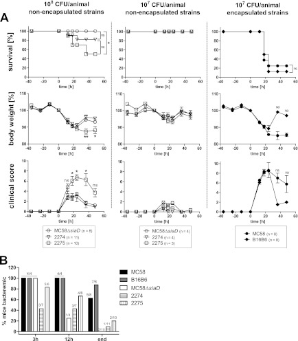
Fig 3.
The mouse intraperitoneal infection model of invasive meningococcal disease. (A) Mice were infected with 107 CFU of encapsulated N. meningitidis strain MC58 or B16B6 (right panels) or with 108 or 107 CFU of N. meningitidis MC58ΔsiaD (capsule-deficient mutant) or the nonencapsulated clinical isolates strain 2274 and strain 2275 (center and left). From 36 h prior to infection until 48 h after infection, mice were monitored every 12 h (and also at 18 h postinfection) for survival (upper panels), loss of body weight (middle panel), and clinical scores, assigned to reflect morbidity (lower panels). Statistic analyses were performed using GraphPad Prism 5. The Mantel-Cox log rank test was used for survival analysis (*, P < 0.05). A one-way analysis of variance applying Bonferroni's post hoc test was used for statistical analysis of body weight (*, P < 0.05, **, P < 0.01). The Kruskal-Wallis test with application of Dunn's post hoc test was used to analyze clinical scoring data (*, P < 0.05). For body weight and clinical score data, only the lowest significance levels of differences between strain 2275 and the other two groups are indicated. na, no statistics applicable because only one (B16B6) or two (MC58) animals survived. (B) Assessment of bacteremia in intraperitoneally infected mice. Tail vein blood samples were drawn at the indicated times after infection of mice and plated to assess viable bacteria in the peripheral blood. Depicted is the percentage of mice testing culture positive for N. meningitidis. Numbers on top of columns indicate the numbers of culture-positive animals among the total number of tested animals.

