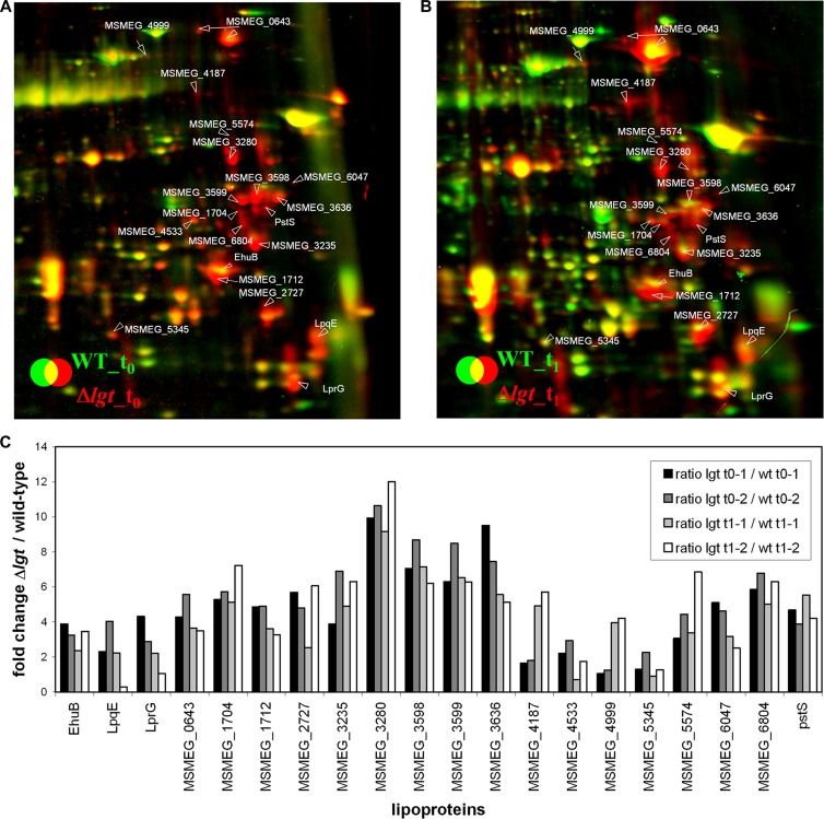
Fig 6.
Close-ups of the lipoproteins that are secreted into the medium in the Δlgt mutant due to the missing lipid anchor. Shown are sections of the dual-channel images of the secretome of the M. smegmatis Δlgt mutant (red image) and the wild-type strain (green image) at the transition phase (t0; A) and 1 h after entry into the stationary phase (t1; B). The secretome was precipitated with TCA and separated using 2D PAGE in the pH range of 4 to 7, as described in Materials and Methods. Quantification of the dual-channel image was performed using Decodon Delta 2D software. (C) The induction ratios of the identified lipoproteins in the extracellular proteome of the lgt mutant to those of the wild type are shown in the corresponding diagram. Two biological replicates are used for quantification in panel C. Proteins are annotated with their corresponding MSMEG_ number. Lipoproteins were identified using LipoP 1.0.

