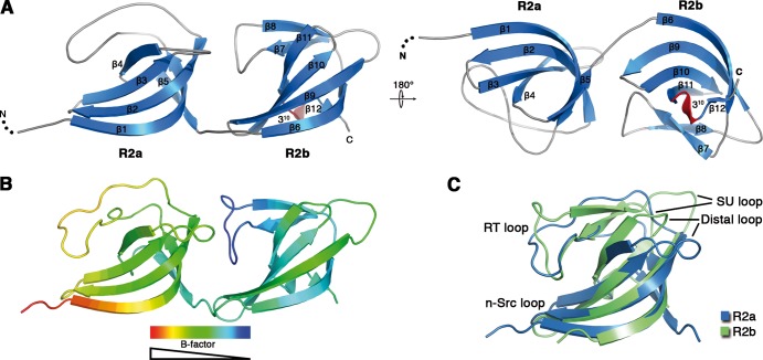
Fig 2.
Crystal structure of the Atl amidase repeat R2ab. (A) Cartoon representation of the crystal structure of R2ab in two orientations. For clarity, loop regions have been smoothed. (B) B-factor distribution in the R2ab structure. B factors decrease from red to blue. (C) Structural comparison of repeats R2a and R2b. The superimposed structures have an RMSD of 2.3 Å (all Cα atoms).

