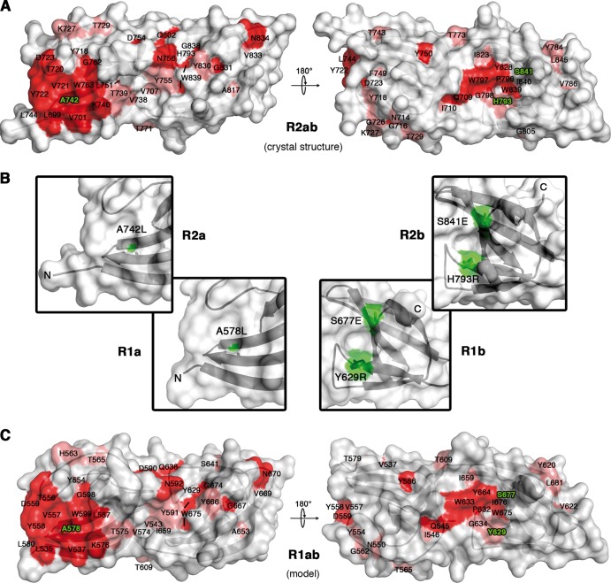
Fig 4.
Conserved residues cluster in two distinct regions on opposite sides of R2ab. (A) Conservation pattern on the surface of R2ab, shown in two different views. Amino acids are colored according to their degree of conservation using the color scheme of Fig. 3. A distinct patch of conserved residues is present on the surface of each subunit in the tandem. Amino acids labeled in green were mutated. R2ab has the same orientation as in Fig. 2. (B) Close-up views of the putative substrate binding sites, with mutated amino acids colored green. (C) Conservation pattern on the surface of R1ab. The R1ab model shown here was generated using MODELLER (12).

