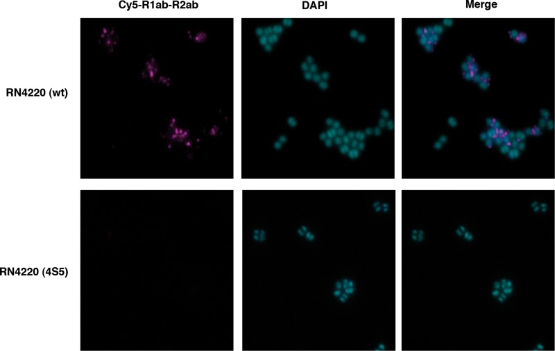
Fig 6.
Distribution of fluorescence-labeled repeats on the surfaces of S. aureus cells. Cells of S. aureus wt and the LTA-deficient strain RN4220 (4S5) were incubated with Cy5-labeled repeats, and the surface distribution was examined by fluorescence microscopy. Chromosomal DNA was visualized by DAPI staining. R1ab-R2ab binding could be observed at the interface between separating cells (top). RN4220 (4S5) did not show fluorescent repeats on the surface (bottom).

