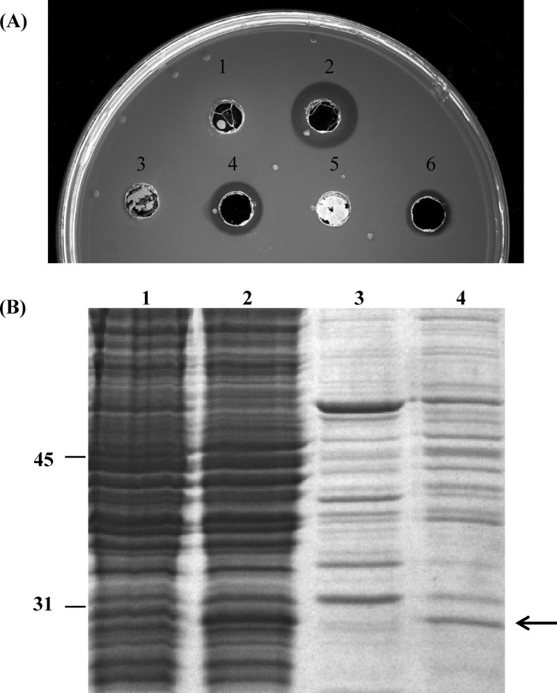
Fig 3.
Cellular location of PhaZBd in E. coli. (A) Enzyme activity measured in mcl-PHA agar plates of cellular fractions: DH10B (pIZ1016) spheroplasts (spot 1); DH10B (pIZBd1) spheroplasts (spot 2); DH10B (pIZ1016) supernatant of the ultracentrifuged periplasmic fraction (spot 3); DH10B (pIZBd1) supernatant of the ultracentrifuged periplasmic fraction (spot 4); DH10B (pIZ1016) pellet of the ultracentrifuged periplasmic fraction (spot 5); and DH10B (pIZBd1) pellet of the ultracentrifuged periplasmic fraction (spot 6). (B) SDS-PAGE analysis of the cellular fractions: lane 1, DH10B (pIZ1016) spheroplasts; lane 2, DH10B (pIZBd1) spheroplasts; lane 3, DH10B (pIZ1016) supernatant of the ultracentrifuged periplasmic fraction; lane 4, DH10B (pIZBd1) supernatant of the ultracentrifuged periplasmic fraction. The arrow shows the position of PhaZBd.

