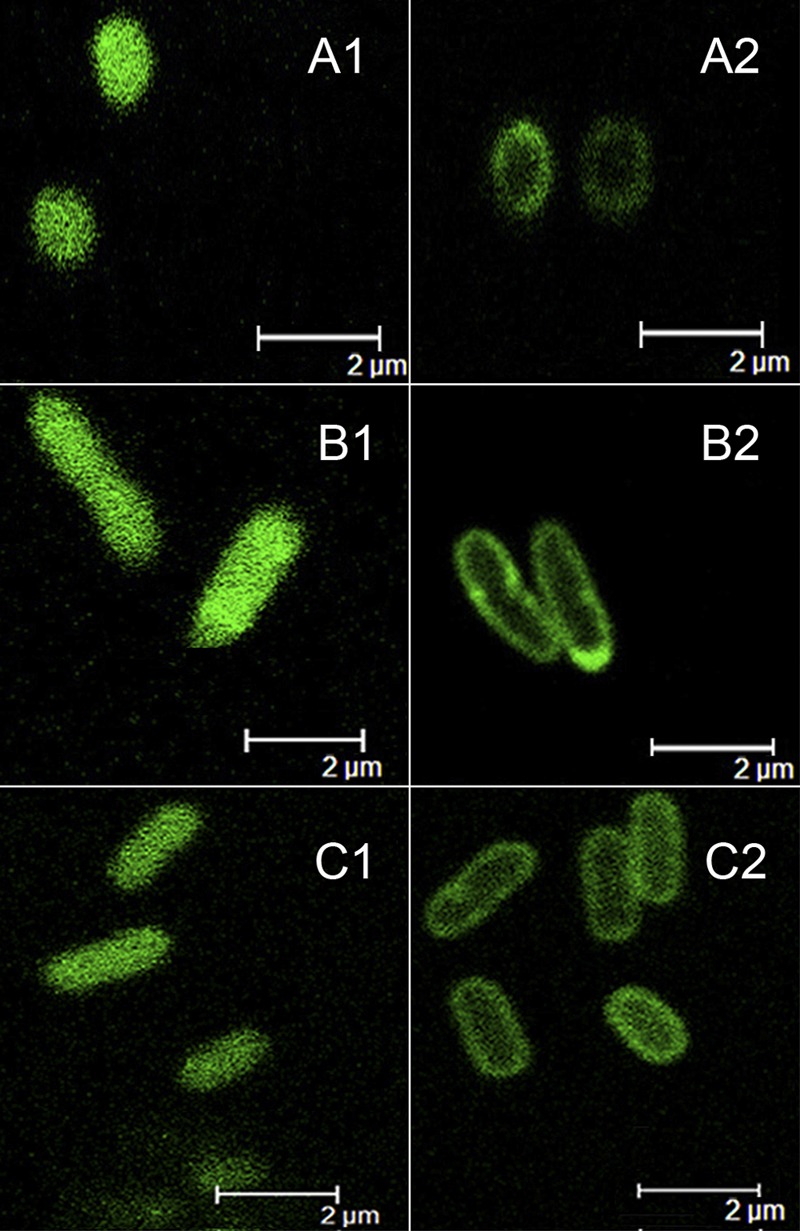
Fig 2.

Localization of green fluorescent proteins by confocal microscopy. (A1, B1, and C1) GFP expressed in K. pneumoniae M5a1[pZWXY005] (A1), P. putida PaW340[pZWXY005] (B1), and E. coli BL21(DE3) harboring plasmid pGFPe (37) (C1) (controls), showing that GFP was distributed throughout the cytoplasm. (A2, B2, and C2) MhbT-GFP expressed in K. pneumoniae M5a1[pZWXY004] (A2), P. putida PaW340[pZWXY004] (B2), and E. coli BL21(DE3)[pZWXY006] (C2), showing that the fusion proteins were all located at the periphery of the cells.
