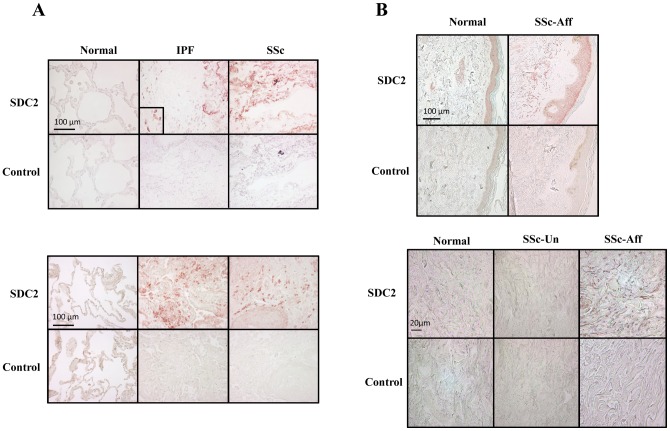
Figure 5. A) SDC2 is highly expressed in fibrotic lung.
Immunohistochemistry was used to detect SDC2 in lung tissues normal donors, patients with IPF, and patients with SSc-associated pulmonary fibrosis. Images are representative of data obtained with lung tissues from 6 normal donors, 9 patients with IPF, and 6 patients with SSc. Rabbit IgG was used as an antibody control. Magnification = 400x. B) SDC2 is over-expressed in SSc affected skin. Immunohistochemistry was used to detect SDC2 in normal donor skin, clinically unaffected and affected skin from a patient with SSc. Images are representative of data from skin of 4 patients with SSc and two controls. Magnification = 200x (left panel), 400x (right panel).

