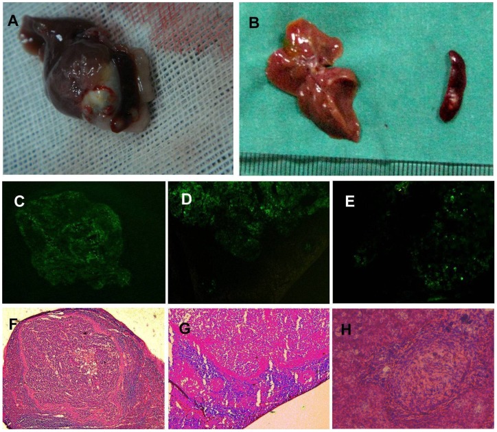
Figure 3. Experimental metastasis model of intrasplenic inoculation into nude mice.
A: Spleen tumors and liver metastases in a macroscopic specimen of the L-SHH group. B: Spleen tumors and liver metastases in a macroscopic specimen of the L-C group. C, D, and E: Fluorescence microscopy images. F, G, and H: Lightmicroscopy images. (C, F: Spleen tumors from the L-Gli1i group; D, G: Intrasplenic miniature metastases from the L-C group; E, H: Liver metastases from the L-SHH group). *P<0.05, **P<0.01.

