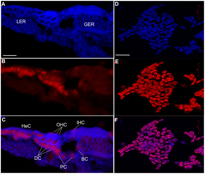
Figure 4. Immunohistochemistry analysis of Hmga2.
Photomicrographs of: (A–C) Cross-sections through the middle turn of the P3 cochlea and (D–F) undifferentiated CGR8 mouse embryonic stem (ES) cells growing in feeder free (leukemia inhibitory factor containing medium) cell culture used as positive control for Hmga2 expression. This positive control was not carried out simultaneously and was only included as a supplemental reference tool. In the P3 cochlea, the expression of the Hmga2 protein (red label) is mostly detected in the supporting cells within the IHC (BC) and OHC (DC) areas, in addition to pillar cells (PC) and Hensen’s cells (HeC). The OHCs are weakly labelled with the Hmga2 antiserum. The ES cells and cross-sections are counterstained with DAPI (shown in blue). IHC: inner hair cells; OHC: outer hair cells; BC: border cells; DC: Deiters’ cells; GER: greater epithelial ridge; LER: lesser epithelial ridge. Scale bars = 25 µm in (A–C) and 50 µm in (D–F).

