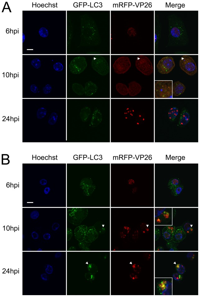
Figure 6. Co-localization of autophagosomes and viral capsids in HSV-1 infected gro29 cells.
L/GFP-LC3 (A) and gro29/GFP-LC3 (B) cells were infected with HSV-1 mRFP-VP26 at an MOI of 10. At 6, 10 and 24 h post infection, the infected cells were fixed and the nuclei were stained with Hoechst. Arrowheads indicate areas in which the mRFP and the EGFP signals co-localize. Insets in the merged panels are magnified regions corresponding to the arrowheads. Confocal images are representative of three independent experiments.

