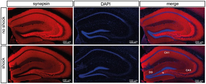
Figure 4. Immunohistochemical analysis of hippocampal synapsin expression.
Representative images (from one mouse out of 6 mice per test condition) of coronal hippocampal sections (bregma −1.6 mm) showing the localization of hippocampal synapsin expression in shocked and non-shocked mice sacrificed 60 days after subjection to footshock (batch PTSD II, behavioral testing d28–30) (left panel). Slices were counterstained with DAPI (middle panel). The overlay of the DAPI and anti-synapsin stained sections is presented in the right panel (merge). Abbreviations: Cornu ammonis areas 1 and 3 (CA1, CA3), dentate gyrus (DG), hilus region (hi).

