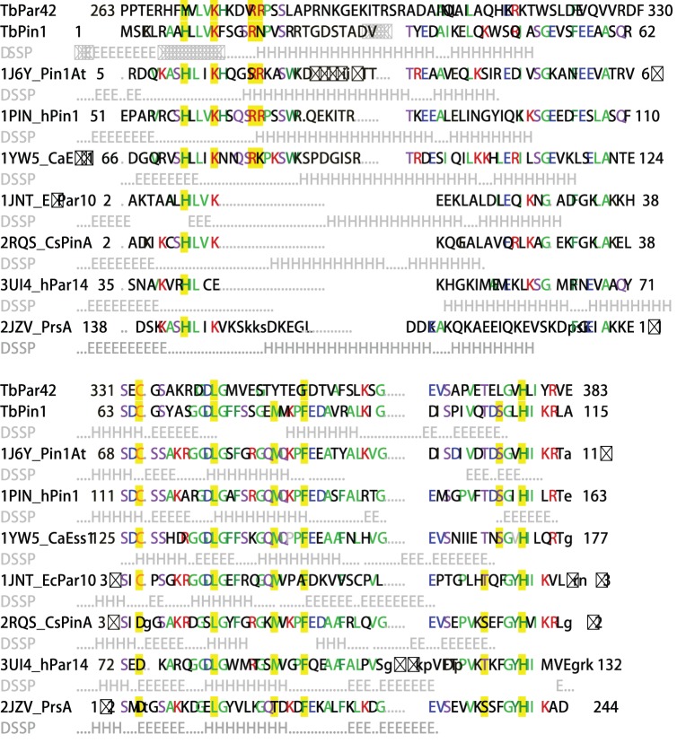
Figure 1. Sequence alignment of TbPin1 with other parvulins.
The following structures and protein sequences were obtained from PDB and SwissProt: TbPin1 (Q57YG1); TbPar42 (Q57XM6); 1J6Y, Pin1At (Q9SL42); 1PIN, hPin1 (Q13526); 1YW5, CaEss1 (G1UA02); 1JNT, EcPar10 (P0A9L5); 2RQS, CsPinA (O74049); 3UI4, hPar14 (Q9Y237); 2JZV, PrsA (P60747). The sequences of parvulins were aligned with that of TbPin1 and the available tertiary structures of parvulins were also aligned onto the structure of TbPin1 using DaliLite [33]. DSSP information for helix (H) and strand (E) conformations from the PDB files is given in light gray below the protein sequences except TbPar42. Aligned residues are written as capital letters and the most frequent residues are colored in each column. Residues considered crucial for the PPIase activity are highlighted in yellow.

