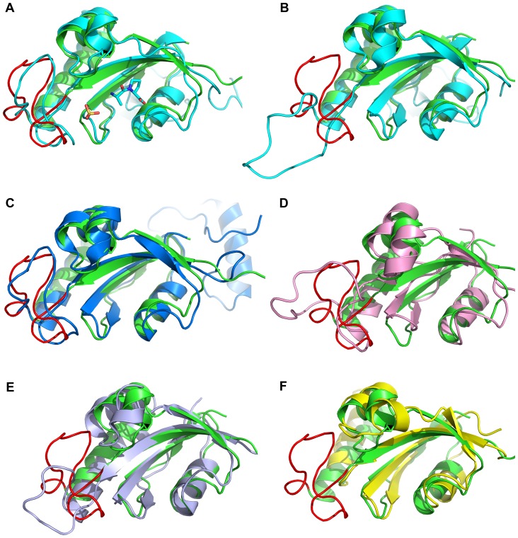
Figure 4. Structural comparison of TbPin1 (2LJ4) with other parvulins.
(A) hPin1 in complex with the Ala-Pro dipeptide and a sulfate ion (1PIN); (B) hPin1 without a sulfate ion (1F8A); (C) CaEss1 (1YW5); (D) Pin1At (1J6Y); (E) PrsA-PPIase domain (2JZV); (F) EcPar10 (1JNT). The structures of TbPin1, hPin1, CaEss1, Pin1At, EcPar10 and PrsA-PPIase domain are colored green, cyan, blue, pink, lightblue and yellow, respectively. The β1/α1 loop of TbPin1 is displayed in red. The sulfate ion is used to mimic to the phosphate group.

