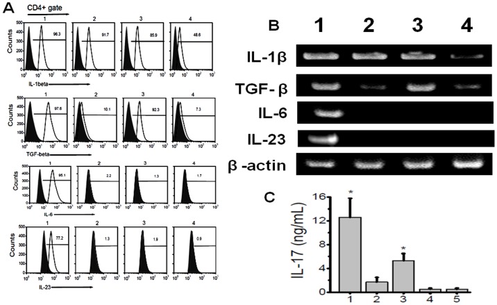
Figure 8. Induction of Th17 cell from naïve T cells by lung infiltrating macrophages.
(A) Flow cytometry analysis for the expression of IL-1β, TGF-β, IL-6 and IL-23 cytokines on CD4+gated population; (B) gene expression of IL-1β, TGF-β, IL-6 and IL-23 in macrophages collected from lungs; (lane1) and peritoneum (lane 3) day 60 MHC class II Ab administered mice, lane 2 and 4 represent macrophages collected from lung and peritoneum of isotype administered animals. (B) represent IL-17 concentration from the media collected by co-culture of 1∶1 ratio of naïve T cells collected from splenocytes of wild type C57BL/6 mice and cultured with macrophages collected from lung (1) and peritoneum (3) of day 60 MHC class II Ab administerd mice, 2 and 4 represent co culture with macrophages collected from lung (2) and peritoneum (4) of day 60 Isotype Ab administered mice, (5 in panel B) represent lung infiltrating macrophages of day 60 MHC class II Ab administered mice in the absence of naïve T cells. The data is represented as a mean ± SEM over a 5 different experiments, the cohort with significant expression of IL-17 (p-value <0.005) compared to isotype Ab administered group (5) were represented with an asterisk (*).

