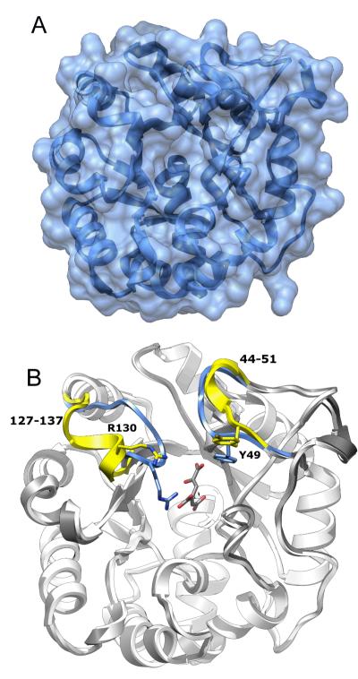
Figure 6.
Structure of native LigI and the D248A mutant. (A) Native LigI. The narrow crevice on the top of the solvent accessible surface corresponds to the active site entrance. (B) View of CHM-bound D248A LigI (white) superimposed onto the wild-type apo-form of LigI (gray). The two loops, Phe-127 to Lys-137 and Ser-44 to Pro-51, that exhibit a change in conformation upon substrate binding, are color coded to the following format: Native LigI (apo) is yellow and CHM-bound D248A is blue.

