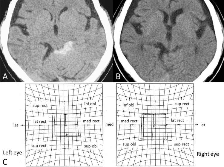
Figure 1.
A) Initial CT. A thick clot is present in the left quadrigeminal subarachnoid space, consistent with perimesencephalic SAH. No oedema or hydrocephalus is observed. B) CT on day 16, showing no SAH in the perimesencephalic region. C) Hess red-green test on day 5. Diplopia is most prominent with right-downward gaze, indicating weakness of the left superior oblique muscle. The Hess chart results are also consistent with left trochlear nerve palsy.

