
FIGURE 6.
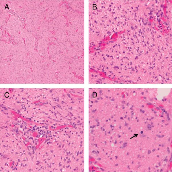
Anaplastic astrocytoma involving the optic nerve. Low-power photograph demonstrates neoplastic glial cells diffusely infiltrating optic nerve fascicles (A; hematoxylin and eosin; 100×). On high power, the tumor cells demonstrate hyperchromasia and marked nuclear atypia (B; hematoxylin and eosin; 400×). Perivascular inflammation was present (C; hematoxylin and eosin; 400×). Mitotic figures were frequent (arrow) (D; hematoxylin and eosin).