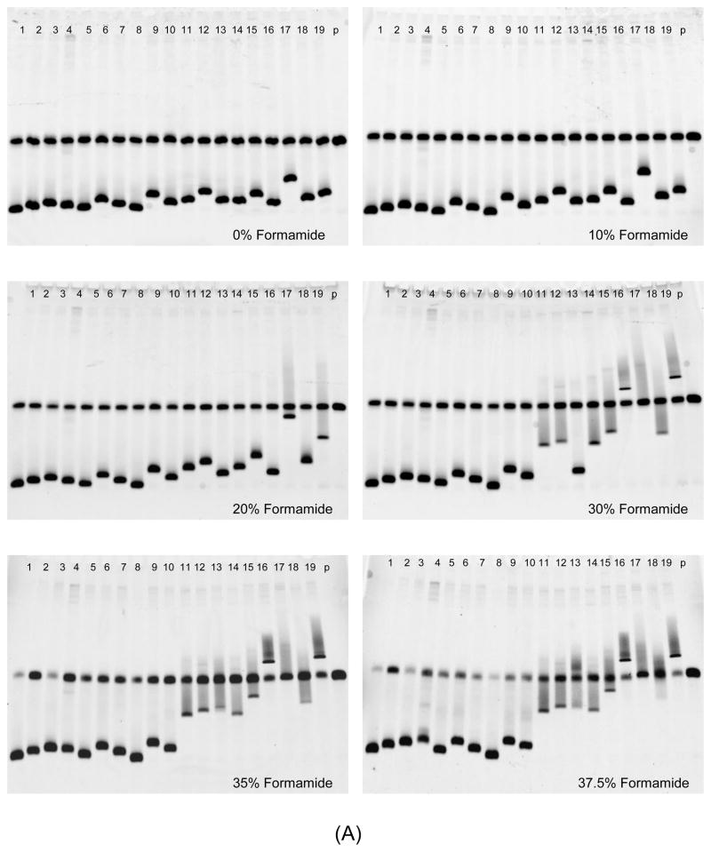
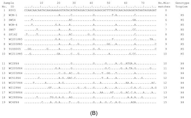
Fig. 3.
Analysis of DNA heteroduplexes with multiple mismatches using HTA with denaturing MDE® gel under different formamide concentrations. (A) HTA gel pictures. Formamide concentration is indicated in each gel. Samples 2 to 19 were heteroduplexes with six to fifteen mismatches, whose sequence alignment is shown as (B). (B) Alignment of target HIV-1 V3 DNA sequences. The sequence starts after primer V3F and ends before primer V3R, corresponding to nucleotide position of HIV-1 V3 11 to 87.


