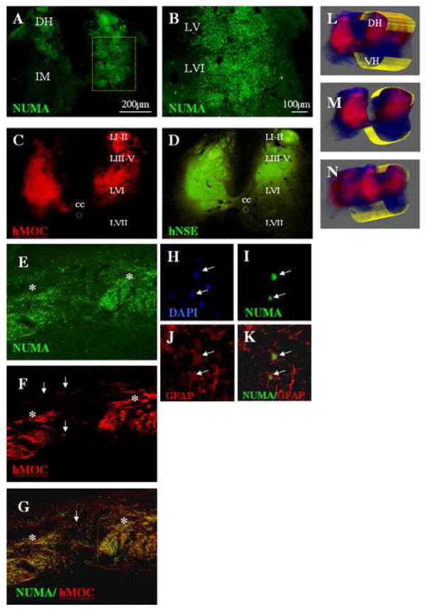
Figure 2 A – N.
A, B: Transverse lumbar spinal cord sections taken at 7 weeks after grafting from an animal injected with 30 000 cells/injection. Staining with human specific nuclear antibody (NUMA) confirmed the presence of grafted cells distributed between the superficial dorsal horn layers and lamina VIII.
C, D: Staining with human specific MOC and NSE antibody revealed intense immunoreactivity within the individual grafts.
E, F, G: Sagittal lumbar spinal cord sections stained with NUMA and MOC antibody. The core of the individual injection sites could clearly be recognized (E, F, G: asterisks). Numerous NUMA- and MOC-positive cells which migrated for distances greater then 500μm from the borders of the grafts can also be identified (white arrows).
H, I, J, K: Confocal analysis of NUMA and GFAP antibody-stained sections showed that a sub-population of NUMA-positive cells differentiated into astrocytes (I, J, K). These cells were typically localized at the periphery of the individual grafts.
L, M, N: Systematic 3-D analysis of serial transverse spinal cord sections stained with the hMOC antibody confirmed a consistent presence of MOC-positive grafts across 3 individual injections sites (DH-dorsal horn; VH-ventral horn).

