
Figure 2. Cathepsin H localization with (Met)enkephalin (ME) in secretory vesicles illustrated by immunofluorescence confocal microscopy of neuronal-like chromaffin cells.
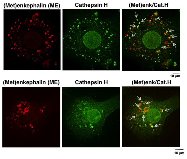
The colocalization of cathepsin H (Cat.H, green immunofluorescence) with ME ((Met)enk, red immunofluorescence) in secretory vesicles is shown by the merged yellow immunofluorescence areas (indicated by arrows) within neuronal-like chromaffin cells. The merged yellow immunofluorescence shows partial localization of cathepsin H with ME. Quantitation of the cathepsin H colocalized with (Met)enkephalin was assessed by measurement of the Pearson’s correlation coefficient (Rr value) of 0.77 + 0.024 (n = 12 cells), which indicates partial colocalization. An Rr value of ‘1’ indicates complete colocalization and a value of ‘0’ indicates no specific colocalization.