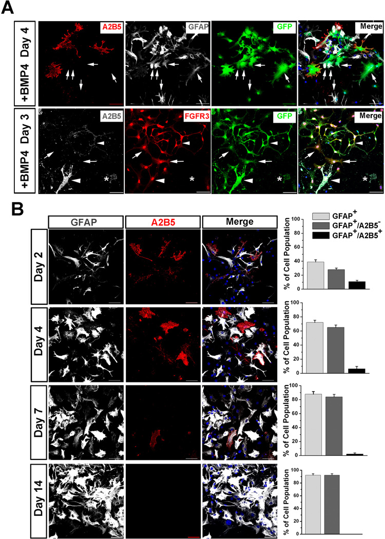
Fig. 3.
Astrocytic differentiation of GFP+ mESC-OPCs in the presence of BMP4. (A) Upper panels after culture in the presence of BMP4, GFP+ mESC-OPCs generate not only GFAP+/A2B5+ type-2 astrocytes, but also GFAP+/− type-1 astrocytes that are indicated by arrows. A2B5 in red, GFAP in white and GFP in green. Lower panelstype-1 astrocytes (indicated by arrows) were also identified by positive for FGFR3 and GFP, but negative for A2B5 staining. A2B5+/FGFR3+ (indicated by arrowheads) and A2B5+/FGFR3− (indicated by star) type-2 astrocytes were identified in the culture. A2B5 in white, FGFR3 in red and GFP in green. (B) Representative images and quantitative analyses showing that the percentage of type-1 astrocytes increases and the percentage of type-2 astrocytes decreases from day 2 to day 7 cultured in the presence of BMP4. A2B5 in red and GFAP in white. Data are presented as mean ± SEM. Blue, DAPI-stained nuclei. Scale bars represent 50 µm.

