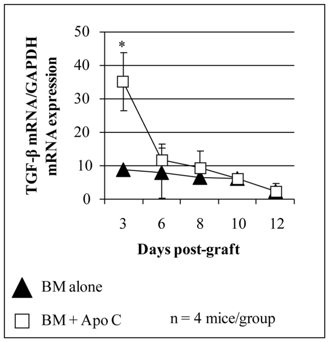
FIGURE 2. Early increase of TGF-β mRNA transcript after intravenous donor apoptotic cell infusion simultaneously with bone marrow graft.

Total RNA was extracted from spleen of BALB/c mice grafted with 106 FVB BM (black triangles) or with 5x10⁶ FVB apoptotic splenocytes (Apo C) plus 106 FVB BM (open square) using RNA extraction kit (Qiagen, Valencia, CA) and reverse transcribed using random hexamers and M-MLV reverse transcriptase (Life Technologies, Rockville, MD) to use as template for quantitative real time RT-PCR (QRT-PCR). QRT-PCR reactions were performed as described (18). Primers and dual labeled fluorescent probes were designed using Primer Express® software (Applied Biosystems, Forster City, CA) to be specific for RNA but not for genomic DNA. QRT-PCR primer pairs and related probes were as follow (sense, antisense and probe respectively). TGF-β : 5′-GCTCTTGTGACAGCAAAGATAACAA-3′, 5′-GGTCGCCCCGACGTTT-3′ and 5′-FAM-CACGTGGAAATCAACGGGATCAGCC-TAMRA-3′; Primer pairs for GAPDH (used as endogenous reference)were already described (18). The relative quantity of each unknown sample was determined automatically with the iCycler iQ® software (Bio-Rad Laboratories, Marnes-la-Coquette, France), using the threshold cycle (Ct). Data were expressed as normalized TGF-β1 expression, which was obtained by dividing the relative quantity of TGF-β1 mRNA for each sample by the relative quantity of GAPDH mRNA of the same sample. *P<0.05.
