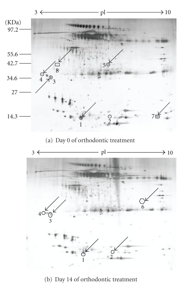
Figure 1.

Representative 2DE gels of human saliva proteome. Saliva proteins resolved by 2-DE (pI 3-10, 24 cm). The resulting gels were visualized by silver staining. Arrows indicate protein spots that changed in expression during orthodontic treatment. The numbers correspond to proteins in Table 1.
