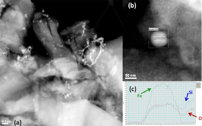
Figure 3.
Transmission electron micrographs of Fe particles on silica gel. (a) Fe particles (~50 nm) formed on surface and in the pores of silica gel. Brighter spots indicate Fe (higher atomic number). Scale bar is 200 nm. (b) Higher resolution image of spherical Fe nanoparticles with path indicating line scan for EDS. (c) EDS confirms elemental Fe composition of particle with silica background.

