
Figure 12.
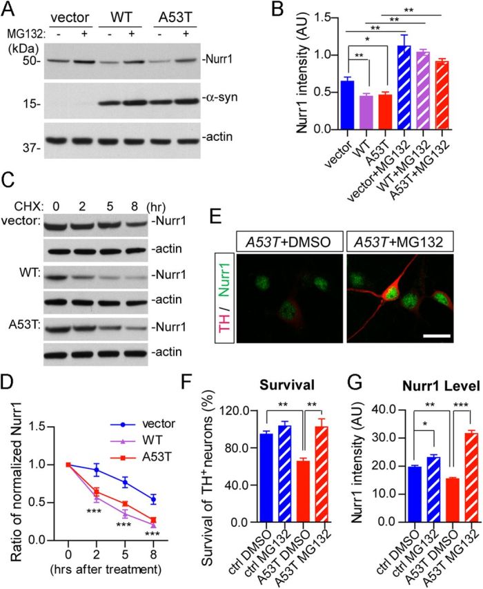
A53T α-syn promotes proteasome-dependent degradation of Nurr1 and inhibition of proteasome activities ameliorates A53T α-syn-induced loss of mDA neurons. A, Expression of endogenous Nurr1 protein in the HEK293 cells transfected with human WT or A53T α-syn cDNA. The transfected HEK293 cells were treated with either DMSO or 10 μm MG132 for 12 h before being lysed for Western blot analysis. β-Actin was used as the loading control. B, The bar graph summarizes the expression of endogenous Nurr1 in α-syn-transfected HEK293 cells treated with DMSO or MG132, from three independent experiments. Data were presented as mean ± SEM. *p < 0.05; **p < 0.01. C, Expression of Nurr1 in human WT or A53T α-syn-transfected HEK293 cells treated with cycloheximide (CHX) for 2, 5, or 8 h before being subjected to Western blot analysis. β-Actin was used as a loading control. D, The line graph traces the decay of endogenous Nurr1 protein in empty vector or α-syn-transfected HEK293 cells from four independent experiments in which CHX was used to block protein synthesis for 2, 5, and 8 h. Data were presented as mean ± SEM. ***p < 0.001. E, Expression of Nurr1 (green) and TH (red) in the A53T midbrain cultures, treated with either DMSO or 10 nm MG132. Scale bar, 10 μm. F, Survival rate (percentage) of TH-positive neurons treated with DMSO or 10 nm MG132. Seven pairs of control and four pairs A53T cultures were analyzed for DMSO or MG132 treatment, respectively. For each culture, 200–500 TH-positive neurons were counted from four independent experiments. Data were presented as mean ± SEM. **p < 0.01. G, Levels of Nurr1 expression in the nucleus of TH-positive neurons in the A53T (n = 145 for DMSO; n = 140 for MG132) and littermate control (n = 117 for DMSO; n = 106 for MG132) cultures treated with either DMSO or MG132. Data were presented as mean ± SEM. *p < 0.05; **p < 0.01; ***p < 0.001.