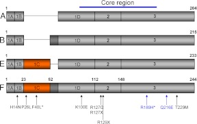
Fig. 1.
Structure of FGF8 isoforms. Schematic representation of the four FGF8 isoforms (A, B, E, and F) resulting from alternative splicing of exons 1C and 1D. The lettering refers to the specific FGF8 isoform. Exons are designated inside the rectangles and numbers above each exon-exon boundary indicate the amino acid position in the FGF8f isoform. The core region of the protein is predominantly made up of exons 2 and 3, which is conserved between isoforms. Previously identified mutations are indicated by arrows and numbered according to the FGF8f protein isoform, with the two novel mutations presented within this paper in blue type. Note that the asterisk denotes homozygous mutations. [Adapted from J. Falardeau et al.: Decreased FGF8 signaling causes deficiency of gonadotropin-releasing hormone in humans and mice. J Clin Invest 118:2822–2831, 2008 (16), with permission. © American Society for clinical Investigation; and E. Trarbach et al.: Nonsense mutations in FGF8 gene causing different degrees of human gonadotropin-releasing deficiency. J Clin Endocrinol Metab 95:3491–3496, 2010 (23), with permission. © The Endocrine Society.

