
Figure 1. Generation of single-cell-derived Pdx1 or Pdx1-VP16-expressing cell clones.
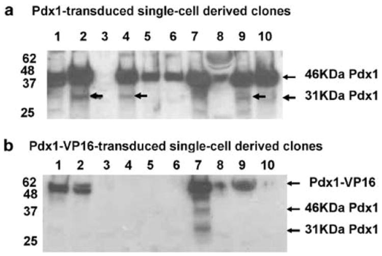
Rat hepatic WB cells were transduced by lentiviral Pdx1- or Pdx1-VP16-vector and single-cell-derived positive clones for either Pdx1 or Pdx1-VP16 were isolated by limiting dilution. The expression of Pdx1 or Pdx1-VP16 was confirmed by separating cellular proteins via SDS-PAGE, transferring to the filter membrane, and blotting with anti-Pdx1 polyclonal antibody (1:5000). (a) Pdx1-tranduced single-cell-derived clones. (b) Pdx1-VP16-transduced single-cell-derived clones. Arrows indicate the positions for active Pdx1 (46 kDa), inactive Pdx1 (31 kDa), and a fusion protein of Pdx1-VP16 (~52 kDa).