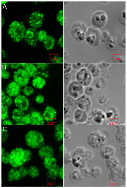
Figure 3.
Immunofluorescence and differential interference (DI) contrast microscopy of P. pastoris overexpressing Ubiquitin. A. 24 h post-methanol induction without nitrogen starvation. B. 48 h post-methanol induction without nitrogen starvation. C. 48 h post-mixed dextrose/methanol induction. Immunofluoresent staining reveals a distribution of Ubiquitin throughout the cytosol and in protein storage bodies. Vacuoles are evident as dark regions in DI contrast images.

