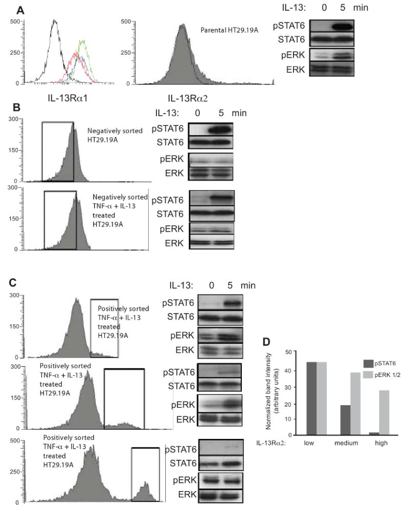
Figure 5. Low IL-13Rα2 expression facilitates ERK activation, while high expression inhibits STAT6.

HT-29.19A cells were treated with IL-13 and TNF-α for 72 h and sorted for IL-13Rα2 expression by flow cytometry generating six groups. Cells were stimulated with 50 ng/ml IL-13. Total cellular proteins were extracted from unstimulated and stimulated cells at 5 min, fractionated by SDS-PAGE, and probed for phospho-specific and total STAT6 and ERK. (Panel A) Untreated or cytokine-treated HT29.19A cells were indirectly immunostained for surface expression of IL-13Rα1 and analyzed by flow cytometry. Isotype control staining is shown in black, parental HT29.19A in red, and the two cytokine-treated cultures in green and blue. Unsorted parental HT29.19A express low amounts of IL-13Rα2. (Panel B) Negatively sorted, untreated or cytokine-treated HT29.19A with no detectable IL-13Rα2 expression. (Panel C) Cytokine-treated, positively sorted HT29.19A for low, medium, or high amounts of IL-13Rα2. (Panel D) Densitometric quantization of the phosphorylated forms of STAT6 and ERK 1/2 normalized to total STAT6 and ERK 1/2 at 5 min of IL-13 stimulation.
