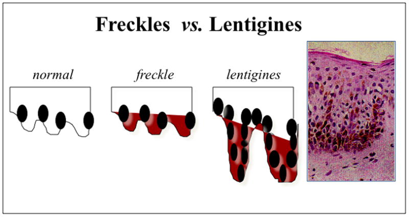
Figure 1.

Histological appearance of freckles vs lenigines Magnification of the lentigen showing melanocytic hyperplasia, characteristic of the lesion (x 200). This differs from common freckles, in which the number of melanocytes is normal but the amount of melanin is increased.
