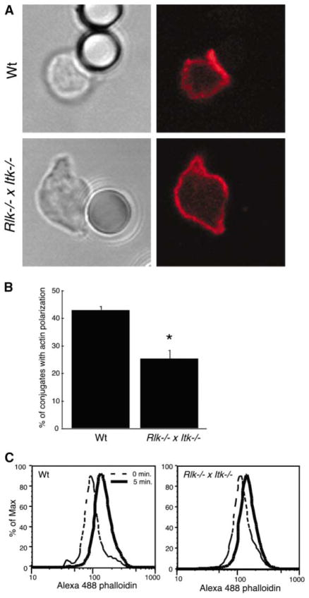
Figure 2. Defects in Actin Polymerization Occur Downstream of TCR Engagement.
(A) Splenic T cells were allowed to conjugate to anti-TCR-coated beads, were fixed, and were stained with Alexa 594-phalloidin to detect F-actin (red). Note the lack of F-actin enrichment at the bead interface with the Rlk−/− × Itk−/− T cell.
(B) Bead conjugates formed as in (A) were scored for accumulation of F-actin. Data are mean ± SD from six independent experiments (the asterisk indicates a significant difference from wt, p < 0.0001).
(C) Splenic T cells were stimulated for 5 min by soluble anti-CD3 crosslinking and were fixed, and F-actin content was assessed after labeling with Alexa 488-phalloidin.

