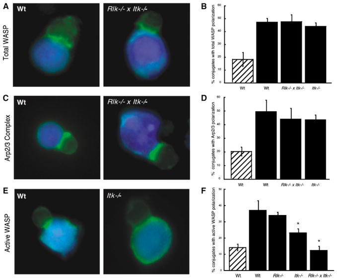
Figure 3. WASP and Arp2/3 Are Recruited to the Immune Synapse, but WASP Remains in an Inactive State.
(A) Conjugates were labeled with an antibody that recognizes WASP in a conformation-independent manner. Both wt and Rlk−/− × Itk−/− T cells show recruitment of WASP to the immune synapse.
(B) Randomly chosen conjugates formed in the absence (hatched bar) or presence (solid bars) of Ag were scored for WASP polarization.
(C) Conjugates were stained with an anti-Arp3 antibody. Both wt and Rlk−/− × Itk−/− T cells show accumulation of Arp2/3 complex at the interface.
(D) Randomly chosen conjugates formed in the absence (hatched bar) or presence (solid bars) of Ag were scored for Arp2/3 polarization.
(E) Conjugates were stained with a monoclonal antibody that preferentially recognizes the open, activated form of WASP. Note that active WASP is not enriched at the immune synapse in the Itk−/− T cell.
(F) Randomly chosen conjugates formed in the absence (hatched bar) or presence (solid bars) of Ag were scored for accumulation of open WASP at the immune synapse.
Data in (B), (D), and (F) represent means from at least three independent experiments ± SD (the asterisk indicates a significant difference from wt + Ag, p < 0.01 for Itk−/−, and p < 0.001 for Rlk−/− × Itk−/− T cells).

