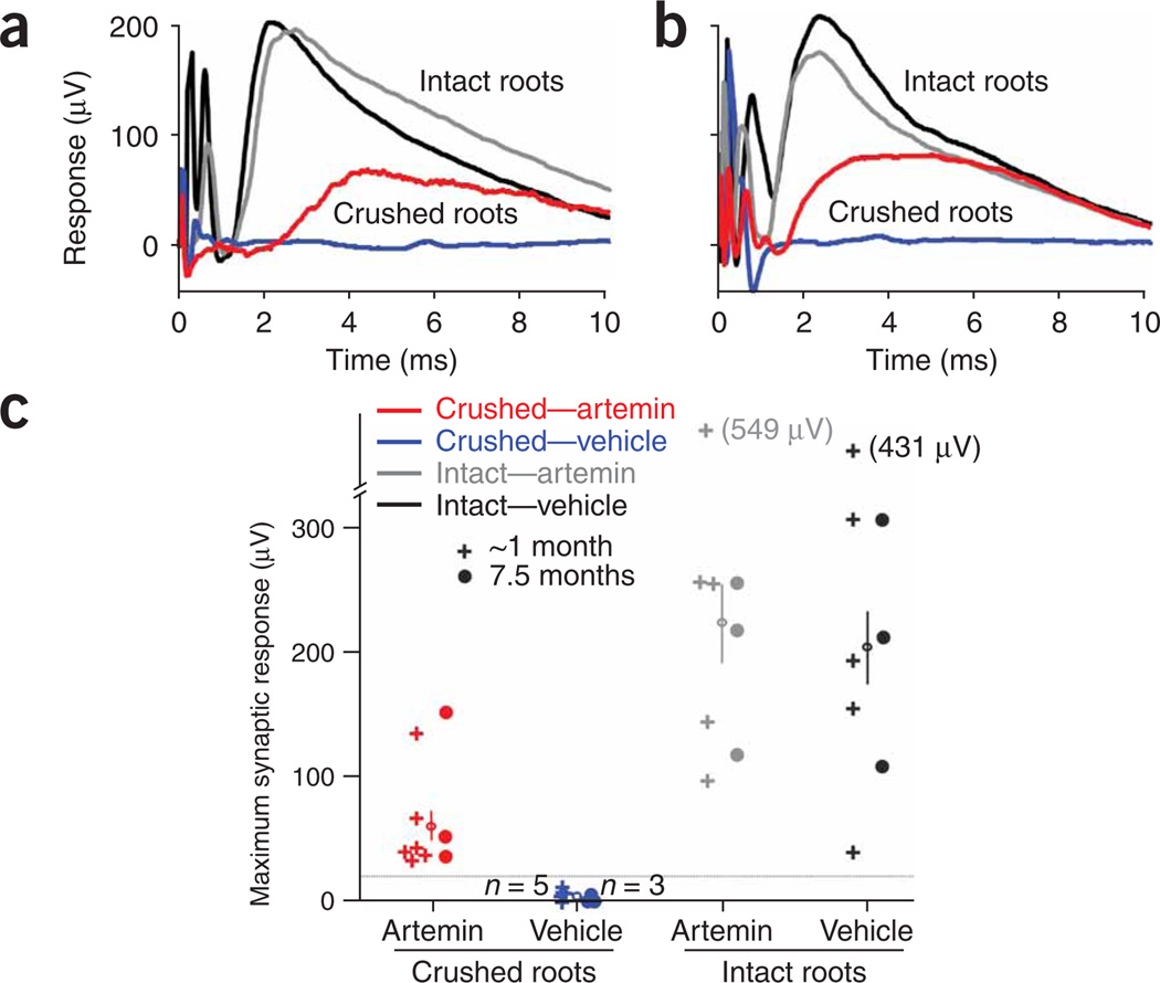
Figure 4.
Systemic artemin administration restores synaptic responses from sensory afferent fibers in spinal neurons. (a,b) Representative traces of field potentials recorded extracellularly in the ventral spinal cord in response to electrical stimulation of the median or radial nerves in the ipsilateral forelimb 1.4 (a) and 7.5 (b) months after DRC. On the unlesioned side of experimental animals (intact roots), the synaptic responses began 1.0–1.5 ms after the stimulus, with rise times of 1.0–1.5 ms in both vehicle-treated (black traces) and artemin-treated (gray traces) animals. In artemin-treated rats, there was substantial recovery of these synaptic inputs (red traces) both 1.4 (a) and 7.5 (b) months after DRC. In contrast, there was no recovery of synaptic function after DRC in vehicle-treated rats (blue traces), even at 7.5 months (b). (c) Scatter plot of the maximum synaptic response to stimulation of the medial or radial nerve recorded in all experimental animals. Each symbol represents the results from one animal, either after DRC or for unlesioned (intact) roots on the contralateral side of the same animal. The average maximum response for each group is shown with an open circle and vertical line (mean ± s.e.m.). The groups tested at ~1 month included postoperative times of 0.7–1.4 months. Colors for the different groups are the same as those used for the traces in panels a and b.

