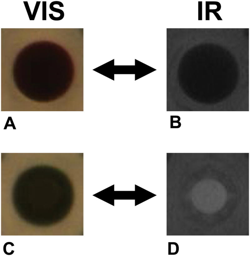
Figure 3. HIAPI-CM distinguishes colored compounds from precipitate.

All images from 384-well microtiter plates. Visible-light images (A, C) of colored compounds can mask the presence of precipitate. However the HIAPI-CM’s IR images of these same wells clearly identifies the top well (images A and B) as containing insoluble precipitate while the bottom well (images C and D) as containing solubilized compound.
