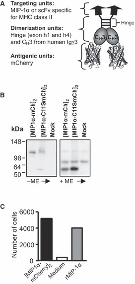
Figure 1.

Characterization of the vaccine constructs containing mCherry. (A) Schematic drawing of the vaccibody protein, which is a homodimer consisting of three functional units: the targeting, the dimerization and the antigenic units. (B) SDS-PAGE (4–20% Tris-Glycine gel) and Western blotting of supernatant harvested from HEK293 transfected with vaccibody DNA. Negative control: supernatant from mock-transfected HEK293. The supernatants were reduced (+ME, mercaptoethanol) or not (-ME) prior to SDS-PAGE. Blotted vaccine proteins were detected by anti-mCherry and chemiluminescence. (C) mMIP-1α in the vaccine format is chemotactic. The indicated vaccibodies were added to the bottom wells of Transwell plates, and the number of CCR1+ CCR5+ Esb-MP cells migrating through the membrane within 2 h was determined by flow cytometry.
