Figure 5.
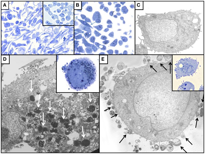
Illustration of the effect of H. pylori colonization on AGS cell monolayers in toluidine blue stained sections [(A,B) inserts of (A,D,E)] and by TEM (C,D,E). (A) WT-infected AGS cells; note the presence of numerous elongated hummingbird cells that are absent among control uninfected AGS cells (insert). (B) AGS cells infected with ΔnudA allele, illustrating that fewer elongated AGS cells (arrows) are present that in WT-infected cells. (C) TEM of uninfected AGS cell with normal ultrastructural aspect. (D) TEM of AGS cell infected with J99 WT H. pylori; note intracellular H. pylori (white arrows). (E) TEM of AGS cell infected with ΔnudA allele; note H. pylori attached to AGS cell (arrows); insert shows the numerous ΔnudA H. pylori attached to AGS cell and relatively few intracellular bacteria. Original magnification of toluidine blue stained pictures (A): 400×; (B) and insert of (A): 1,000×; inserts of (D,E): 1,000×. Original magnification of TEM pictures: (C): 9,800×; (D): 32,500×; (E): 26,000×.

