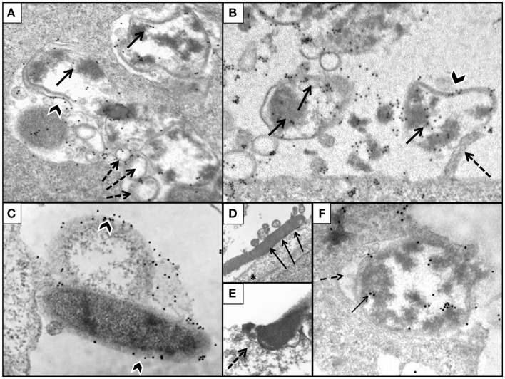
Figure 7.
Illustration of AGS cells with pre-embedded H. pylori immunogold. (A) AGS cell infected with H. pylori WT demonstrating intracellular presence of bacteria located inside vacuoles (gold marked bacterial wall, black arrowhead); note gold particles tagging H. pylori cytoplasm (arrow) and spherical membrane vesicles (dashed arrows). (B) ΔnudA allele with numerous bacteria labeled with gold (arrows) in close association with cell membrane; one of the bacteria is attached to cell pedestal (dashed arrow). (C) Mature and coccoid ΔnudA allele in close association with AGS cell membrane; note bacterial membrane is tagged with gold (arrowheads). (D) ΔnudA allele in early stage of adhesion to AGS cell; note multiple fusions between membranes of bacterium and AGS cell (arrows). (E) Higher magnification of “zipper-like” ΔnudA allele allele entry into AGS cell; note “cup formation” (dashed arrows) identifying H. pylori and AGS cell membranes fusing together during internalization. (F) ΔnudA allele almost completely englobed within invagination of AGS cell membrane; note that this fusion is accompanied by the presence of filamentous strands (fibrils), dense round spheres, vesicles, and amorphous deposits in the space between the membranes of the bacterium and AGS cell (dashed arrow). Immunogold-tagged bacterium (solid arrow) Original magnification (A–C): 41,000×; (D): 24,500×; (E): 32,500×; (F): 41,000×.

