
Figure 7.
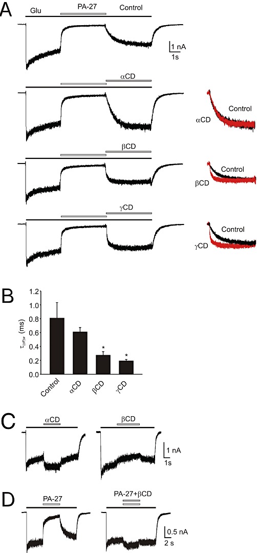
Cyclodextrin effects on the rate of recovery from PA-27-induced inhibition. (A) Example of a response to 1 mmol·L−1 glutamate and its co-application with 10 µmol·L−1 PA-27 (Control), and that recorded in the presence of 10 mmol·L−1α-cyclodextrin (αCD), 10 mmol·L−1β-cyclodextrin (βCD) or 10 mmol·L−1γ-cyclodextrin (γCD). On the left, recovery recorded in the absence of cyclodextrin (Control) and that recorded in the presence of cyclodextrin (in red) are shown normalized and superimposed. Note that the rate of recovery from PA-27-induced inhibition was accelerated in the presence of βCD and γCD. All records are from the same cell. (B) Summary of the effects of α-, β- and γ-cyclodextrin on the rate of recovery from inhibition induced by PA-27. Abscissa shows mean ± SD of the weighted single or double exponential fit to the recovery after steroid application (n= 5). *P < 0.05, significantly different from Control, paired t-test. (C) Effect of 10 mmol·L−1α- and β-cyclodextrin on 1 mmol·L−1 glutamate-induced responses. (D) Example of a response to 1 mmol·L−1 glutamate and its co-application with 10 µmol·L−1 PA-27 (Control), and that recorded in the presence of mixture of 10 mmol·L−1β-CD and 10 µmol·L−1 PA-27.