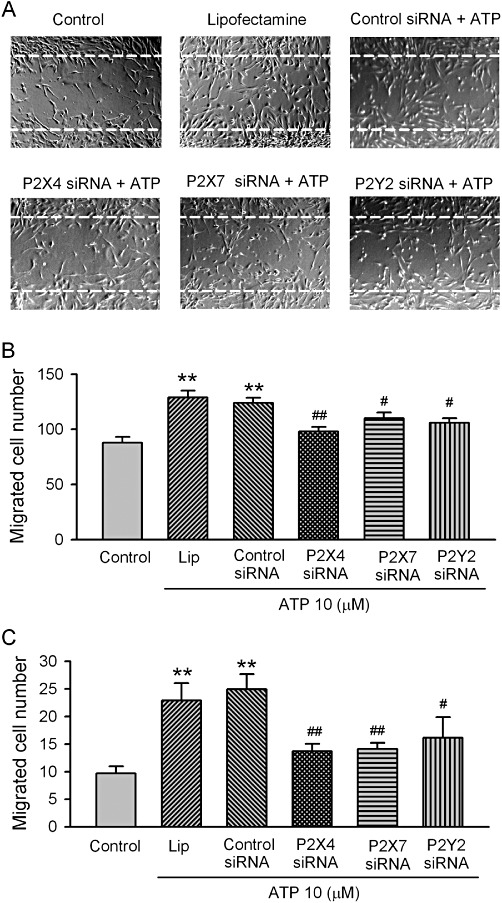
Figure 8.

Effect of ATP on the migration of human cardiac fibroblasts. (A) Images of human cardiac fibroblasts in the wound-healing migration assay. Confluent cardiac fibroblasts were scraped off with a pipette tip to induce acellular areas, then treated 10 µM ATP in cells transfected with Lipofectamine, control siRNA, P2X4, P2X7 and P2Y2 siRNAs, respectively (40 nM each). Images were taken after 20 h incubation with 10 µM ATP. Broken white lines indicate the initial acellular wound regions. (B) Mean values for number of migrated human cardiac fibroblasts counted in areas as marked in (A) (n= 4, **P < 0.01 vs. control, #P < 0.05, ##P < 0.01 vs. control siRNA). (C) Mean values for number of migrated human cardiac fibroblasts counted on lower surface of the Transwell membrane (microchemotaxis assay with 10 µM ATP incubation for 6 h, n= 3, **P < 0.01 vs. control, #P < 0.05, ##P < 0.01 vs. control siRNA).
