
Figure 3.
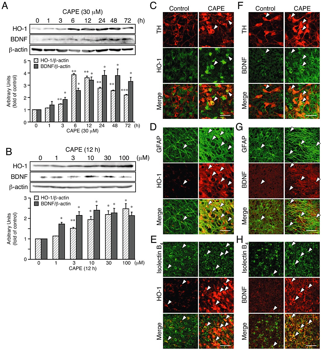
CAPE increases expression of HO-1 and BDNF in midbrain slice cultures. (A, B) Western blot analysis of the effect of CAPE on expression levels of proteins for HO-1 and BDNF. Six slices for each condition were pooled as sample for each lane. Midbrain slice cultures were treated with 30 µM CAPE for indicated periods (A) or with CAPE (1–100 µM) for 12 h (B). n= 3. *P < 0.05, **P < 0.01, ***P < 0.001 versus corresponding control (anova results: for HO-1 in panel A, F(7,16) = 223.64, P < 0.0001; for BDNF in panel A, F(7,16) = 12.92, P < 0.0001; for HO-1 in panel B, F(5,12) = 17.36, P < 0.0001; for BDNF in panel B, F(5,12) = 9.23, P= 0.0008). (C–H) Confocal images of double immunofluorescence histochemistry of cell type markers as indicated (upper panels) with HO-1 (C–E) and BDNF (F–H), respectively (middle panels), and their merged images (lower panels) in control cultures (left panels) and cultures treated with 30 µM CAPE for 12 h (right panels). Representative double-positive cells are indicated by arrowheads. Scale bars, 50 µm. Cell type markers are TH (dopaminergic neurons), GFAP (astrocytes) and isolectin B4 (microglia).