
Figure 4.
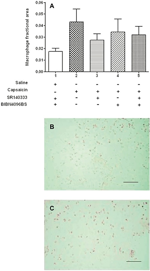
Detection of macrophages in the synovium. (A). Low levels of macrophage infiltration in all groups. There were no statistically significant differences in macrophage fractional areas for each group of rats, indicating that any small cellular inflammatory response present was below the detection threshold. Data shown are means ± SD; n = 6 rats per group Macrophages in the synovium were detected by immunohistochemistry using the primary antibody ED1 and were stained with nickel/DAB and appear as black. (B) Synovium from a capsaicin-treated animal. (C) Synovium from a saline-injected knee of an animal treated with the CGRP receptor antagonist BIBN4096BS i.v. and the SP receptor antagonist SR140333 i.p. The number of positively staining cells in each panel appears similar. When quantified, there were no statistically significant differences in the macrophage fractional area between any of the groups. Bar = 100 µm.