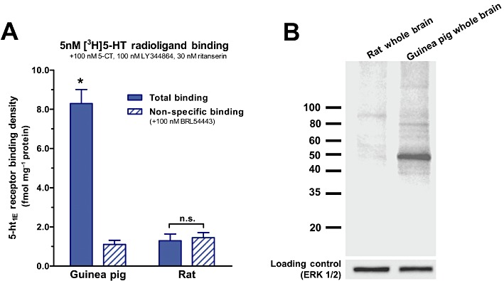
Figure 1.

Specificity of the anti-5-ht1E antibody assessed by Western blot. (A) [3H]5-HT radioligand binding demonstrates that 5-ht1E receptors are expressed in guinea pig brain tissue (*P < 0.001, two-way anova with Bonferroni corrected pair-wise comparison) but not in rat brain tissue. Drugs are present at concentrations to mask [3H]5-HT binding to non-5-ht1E receptor sites (Klein and Teitler, 2009). Data are the means ± SEM of three independent experiments performed in triplicate. (B) Western blot analysis of guinea pig and rat whole brain lysates agrees with radioligand binding: the anti-5-ht1E antibody intensely stains a single ∼45 kDa band in guinea pig but not rat brain lysates. The predicted molecular weight of the 5-ht1E receptor is 43 kDa. The immunoblot is representative of three independent experiments.
